Eye pressure can matter in emergency care. But does measuring it need to require a traditional ophthalmic workflow or specialized knowledge of the cornea?
Patients with eye pain, redness, visual changes, headache, nausea, trauma, and other ocular complaints routinely present to emergency departments and urgent care centers.
Some require immediate ophthalmologic evaluation. Others require clinicians to rapidly distinguish potentially vision-threatening conditions from less urgent problems.
Intraocular pressure (IOP) can be an important part of that assessment.
Yet obtaining and interpreting IOP outside a dedicated ophthalmology or optometry practice can introduce practical challenges.
The equipment may not be immediately available. The patient may be lying on a stretcher. Staff may have limited experience with ophthalmic instruments. Some tonometry methods require corneal contact, topical anesthetic, disposable probes or covers, or additional equipment.
There is another issue that receives less attention:
What does the clinician know about the patient’s cornea?

When IOP is measured through the cornea, the cornea itself can influence the measurement.
That may be routine knowledge in an eye-care practice. In an emergency department or urgent care center, however, detailed corneal history, pachymetry, and specialized corneal assessment may not be readily available.
This raises an important question:
What if you take the cornea out of the equation?
When IOP information matters, how easily can your team obtain it?
Tonometry is not necessary for every patient presenting with an eye complaint.
However, there are clinical situations in which IOP becomes an important part of the overall evaluation.
Acute angle-closure glaucoma is one obvious example. Patients can present with severe eye pain, headache, blurred vision, halos, nausea or vomiting, conjunctival injection, and corneal changes. Markedly elevated IOP is one of the characteristic findings requiring urgent evaluation and management.
IOP may also contribute useful information in the evaluation of other ocular presentations.
At the same time, tonometry must be performed appropriately. For example, when an open-globe injury is suspected, pressure should not be applied to the eye and tonometry is generally avoided.
The question therefore isn’t whether every emergency or urgent-care patient should undergo tonometry.
The more practical question is:
When IOP information is clinically indicated, how readily can your team obtain it?
Tonometry involves more than owning a tonometer
Consider the workflow associated with some traditional corneal-contact methods.
Depending on the technology, obtaining an IOP measurement may involve topical anesthetic, disposable probe covers or probes, positioning requirements, direct corneal contact, calibration procedures, and staff training.
None of those requirements makes a technology inappropriate. In an ophthalmology practice, they may simply be part of the normal workflow.
But emergency medicine and urgent care operate differently.
The patient may be on a stretcher.
The clinician may be moving between multiple treatment rooms.
Ophthalmology may not be immediately available.
The staff member performing the measurement may not routinely perform comprehensive eye examinations.
Supplies need to be available when needed.
And the instrument needs to fit into an already busy clinical workflow.
Therefore, evaluating tonometry for an ED or urgent care center shouldn’t begin only with:
“Which tonometer should we buy?”
It should also ask:
“What does it actually take for our staff to obtain a useful IOP measurement?”
The hidden variable: the cornea
There is another consideration that goes beyond workflow.
Many commonly used tonometry technologies obtain IOP information through interaction with the cornea.
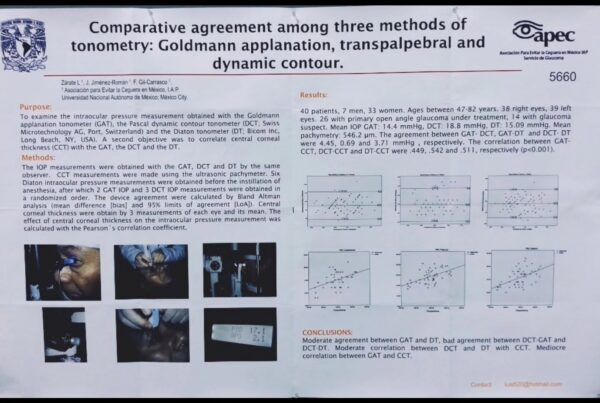
Goldmann applanation tonometry, for example, estimates IOP based on the force required to flatten a defined area of the cornea. It remains a clinical reference standard, but its measurements can be influenced by characteristics of the cornea itself.
Central corneal thickness is one well-recognized variable.
In general, unusually thin corneas may result in lower measured IOP with some corneal-dependent methods, while thicker corneas may result in higher measurements.
But thickness is only part of the story.
Corneal biomechanics, curvature, previous refractive surgery, ectasia, edema, scarring, irregularity, and other changes can also influence corneal-dependent tonometry and its interpretation.
This means that a seemingly simple number can contain another question:
How much is the cornea affecting your IOP reading?
A pressure of 17 mmHg may not tell the entire story
Imagine an emergency department patient whose IOP is measured at 17 mmHg.
The number by itself may appear unremarkable.
But consider what may not be known at the time of measurement:
Has this patient had LASIK or PRK?
Do they have an unusually thin or thick cornea?
Could they have keratoconus or another corneal ectasia?
Is the cornea edematous?
Is there significant corneal scarring?
Has the patient undergone previous corneal surgery?
Are the corneal biomechanics otherwise altered?
The emergency clinician may not know.
The patient may not know.
And even if the patient knows, they may not think to mention a refractive procedure performed 15 or 20 years earlier while being evaluated for an unrelated acute complaint.
That does not mean a corneal-dependent IOP measurement has no clinical value.
It means the condition of the cornea is part of the context in which that measurement should be interpreted.
LASIK is a good example
Millions of patients have undergone LASIK, PRK, and other corneal refractive procedures.
These procedures intentionally alter corneal anatomy and biomechanics.
AAO educational material notes that changes following LASIK can produce artifactually low IOP measurements with Goldmann applanation tonometry. Similar concerns exist following other corneal refractive procedures.
In an ophthalmology office, that history may already be documented and incorporated into clinical interpretation.
In an emergency department or urgent care center, it may not be.
This illustrates a larger issue:
When the measurement passes through the cornea, understanding the number may require understanding the cornea.
Does pachymetry solve the problem?
Pachymetry provides valuable information about central corneal thickness and has an important role in ophthalmic care.
But CCT is not the same thing as complete corneal characterization.
Corneal biomechanics are complex. Thickness alone does not fully describe how a particular cornea behaves during a pressure measurement.
There is also no universally accepted mathematical formula that simply converts every corneal-dependent IOP measurement into a definitive “true IOP” based on CCT alone.
So even if pachymetry is available, another question remains:
Is the cornea otherwise normal?
For an ophthalmologist or cornea specialist, evaluating that question is part of specialized training.
For an emergency physician, urgent-care clinician, nurse, technician, internist, or other healthcare professional outside dedicated eye care, detailed corneal assessment is understandably not their primary specialty.
And it shouldn’t necessarily have to be.
What if measuring IOP didn’t require interpreting the cornea first?
This is where a different tonometry approach becomes interesting.
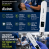
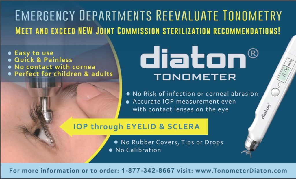
Diaton does not obtain its IOP measurement through the cornea.
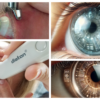
Instead, the device measures transpalpebrally through the upper eyelid over the scleral region.
The FDA-cleared Diaton Tonometer is intended for measuring intraocular pressure as an aid in the diagnosis of glaucoma and for monitoring IOP. Its FDA documentation describes measurement through the eyelid in the scleral area.
Because the cornea is not the measurement surface, central corneal thickness is not an input required to obtain the Diaton IOP measurement.
And the operator does not need to determine whether LASIK, keratoconus, corneal thickness, edema, scarring, or another corneal characteristic is influencing a corneal-based measurement, because Diaton isn’t measuring through the cornea in the first place.
That’s an important distinction.
Diaton does not eliminate the need to recognize or treat corneal disease.
A patient with keratoconus, corneal edema, an abrasion, infection, trauma, or other pathology may still require appropriate ophthalmic examination, diagnosis, and treatment.
Diaton simply removes the cornea from the IOP measurement itself.
This could matter particularly outside ophthalmology
Consider the difference between an ophthalmology office and an urgent care center.
An ophthalmology practice may already have:
specialized eye-care personnel,
slit lamps,
pachymetry,
corneal topography or tomography,
complete ophthalmic history,
multiple tonometers,
and clinicians trained to recognize subtle corneal abnormalities.
An urgent care center or emergency department may not.
That isn’t a deficiency.
They’re designed to do different jobs.
The question is whether obtaining useful IOP information should necessarily require the same infrastructure and corneal expertise available in a specialized eye clinic.
A cornea-independent measurement approach potentially changes that equation.
Bringing IOP measurement to the bedside
Diaton’s potential value in emergency medicine isn’t limited to its relationship with the cornea.
Its FDA documentation describes a compact handheld device that can be used with appropriately positioned patients in sitting or supine positions and does not require topical anesthesia for its measurement.
That creates a different workflow proposition:
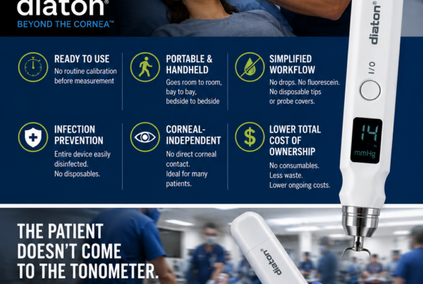
No corneal contact
No topical anesthetic required for the Diaton measurement
No disposable corneal probe or tip cover for each measurement
No CCT input required to obtain the Diaton reading
Portable
Sitting or supine measurement
No slit lamp required to obtain the measurement
Each is a product characteristic.
But together they potentially provide something more important to an ED or urgent care center:
Greater practical access to IOP measurement.
Should the measurement come to the patient?
Emergency medicine has progressively moved diagnostic capabilities closer to patients.
Blood pressure is measured wherever the patient is.
Pulse oximetry travels with the patient.
Electrocardiography comes to the bedside.
Point-of-care ultrasound has increasingly brought imaging directly into emergency examination and resuscitation environments.
Perhaps IOP measurement should be considered through a similar operational lens.
Instead of asking:
“How do we get this patient to the eye equipment?”
we can ask:
“How do we bring IOP measurement to this patient?”
A handheld approach that can be used with a sitting or appropriately positioned supine patient may fit naturally into that model.
The goal isn’t to turn emergency clinicians into ophthalmologists
This distinction is critical.
Diaton does not diagnose glaucoma by itself.
An IOP number alone does not determine the diagnosis or management of an ocular emergency.
And a tonometer does not replace an ophthalmologic examination when one is clinically indicated.
Rather, IOP is another piece of clinical information.
The objective is not to ask emergency physicians, urgent-care clinicians, nurses, or technicians to become corneal specialists.
In fact, that is part of the point.
Obtaining a Diaton IOP measurement does not require the operator to first characterize the patient’s cornea.
The measurement follows another anatomical route.
When something looks wrong, when IOP is concerning, or when the overall clinical presentation warrants specialist care, the patient should still be appropriately escalated or referred.
The tonometer provides information.
The clinician provides judgment.
Think about the entire IOP workflow
When an emergency department, urgent care organization, or hospital evaluates tonometry equipment, purchase price is only one consideration.
Decision-makers should consider the entire process:
Who will perform the measurement?
Where will it happen?
Can the device come to the bedside?
Can it be used with a patient who is supine?
Does it require corneal contact?
Are anesthetic drops required?
Are disposable probes or covers required for each measurement?
Does interpretation depend on corneal characteristics that may be unknown?
Is pachymetry needed as part of the measurement workflow?
Does staff need specialized knowledge to recognize when corneal characteristics may be influencing the measurement?
What training is required?
What does the equipment cost?
What does every subsequent measurement cost?
And ultimately:
Will the device actually be used when IOP information is needed?
That last question may matter more than almost any specification on a product sheet.
From buying equipment to building IOP capability
Hospitals and urgent care centers don’t ultimately need another piece of equipment sitting in a cabinet.
They need clinical capability.
An effective IOP program should therefore consider appropriate indications and contraindications, staff training and competency, infection-control procedures, documentation, device availability, and a defined pathway for ophthalmologic escalation when appropriate.
This is also where implementation support matters.
Providing a portable tonometer without ensuring that clinicians are comfortable using it does not solve the underlying problem.
The objective should be to create an environment in which appropriately trained personnel can obtain IOP information when clinically indicated and understand when specialist evaluation is necessary.
A different way to evaluate tonometry outside eye care
For decades, tonometry has largely been viewed as ophthalmic equipment.
But perhaps hospitals and urgent care organizations should evaluate the problem differently.
Not:
Which ophthalmic tonometer should we own?
But:
What is the simplest appropriate way to give our clinicians access to IOP information when they need it?
That changes the conversation.
It shifts the emphasis from a device specification sheet toward:
Access
Workflow
Training
Portability
Corneal independence
Cost per measurement
Clinical implementation
And importantly:
It removes the assumption that obtaining an IOP measurement has to begin with the cornea.
Think Beyond the Cornea.™
When the cornea is the measurement surface, its characteristics can become part of the number.
When those characteristics are unknown, interpretation may become more complicated.
Diaton provides another approach.
IOP through the eyelid and sclera.
No corneal contact.
No CCT input required to obtain the measurement.
No need to determine whether corneal characteristics are affecting a corneal-based measurement, because the measurement doesn’t pass through the cornea.
For emergency departments, urgent care centers, hospitals, and other settings outside dedicated eye care, that may represent more than another way to measure pressure.
It may represent a more practical way to make IOP measurement accessible where the patient already is.
Think Beyond the Cornea.™
Evaluating IOP measurement for your Emergency Department or Urgent Care Center?
Diaton offers hospitals and urgent care organizations a different approach to bedside IOP measurement.
Request information about a Diaton Hospital & Emergency Medicine Evaluation.
Learn more about Diaton Tonometer:
Glaucoma screening / glaucoma awareness
100 Questions and Answers on Diaton
How to use the Diaton Tonometer technique
Diaton in post-LASIK / corneal pathology
Diaton in emergency medicine / broader screening environments